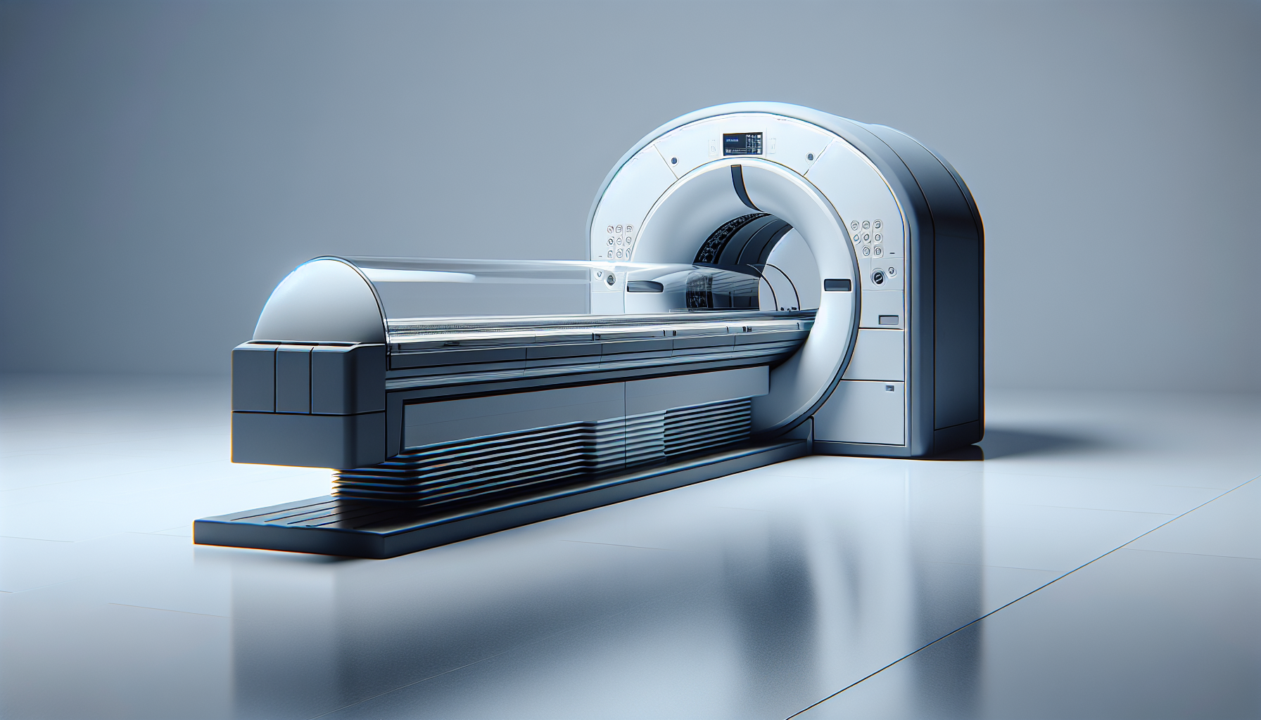
Have you ever wondered what the acronym PET stands for? Well, you’re in luck! In this article, we’ll explore the full meaning of PET and uncover the fascinating world behind this three-letter abbreviation. Whether you’ve encountered it in a medical context, environmental discussions, or even in the realm of fashion, it’s time to uncover the true meaning of PET and unravel its versatile nature. So, let’s dive in and discover what lies beneath the surface of this intriguing acronym.
What is PET?
Introduction to PET
PET, which stands for Positron Emission Tomography, is a powerful imaging technique used in the medical field to obtain detailed images of the inside of the body. It is a non-invasive procedure that involves the use of radioactive tracers and a specialized scanner to detect and visualize cellular and molecular activity. PET scans provide valuable information about various diseases and conditions, aiding in diagnosis, staging, and treatment monitoring.
Definition of PET
Positron Emission Tomography, or PET, is a medical imaging technique that utilizes radioactive isotopes to produce images of the body’s metabolic and physiological processes. By injecting a small amount of a radioactive substance, known as a radiotracer, into the patient’s bloodstream, PET scans can capture the distribution and concentration of the tracer within the body. The emitted positrons from the radiotracer’s decay are detected by the PET scanner, which generates detailed three-dimensional images of the targeted area.
Importance of PET
The significance of PET lies in its ability to provide a unique perspective on the inner workings of the human body. Unlike other imaging modalities, PET focuses on functional processes, offering valuable information about cellular metabolism and physiological functions. This enables physicians to detect diseases in their early stages, determine the extent of the disease, assess treatment efficacy, and guide surgical interventions. PET has become an invaluable tool in various medical specialties, including oncology, neurology, cardiology, and pharmaceutical research.
History of PET
Early Development of PET
The origins of PET can be traced back to the 1950s when scientists Herbert L. Wagner and Jens C. Egeving developed the theory and principles behind this groundbreaking imaging technique. In the early stages, PET was limited to experimental research due to the complexity of equipment and lack of suitable radiotracers. However, pioneering efforts by Michael E. Phelps and his team in the 1970s paved the way for the clinical application of PET by introducing new radiopharmaceuticals and developing sophisticated image reconstruction algorithms.
Advancements in PET Technology
Over the years, PET technology has undergone significant advancements, leading to improved imaging quality and diagnostic accuracy. The development of hybrid imaging systems, such as PET/CT and PET/MRI, revolutionized the field by combining the functional information from PET with the anatomical details provided by CT and MRI. Additionally, the introduction of time-of-flight PET scanners and newer detector materials has enhanced image resolution, reducing scan time and improving overall patient experience.
Impact of PET on the Medical Field
PET has had a profound impact on the medical field, transforming the way diseases are detected and managed. In oncology, PET scans play a vital role in cancer diagnosis, staging, and treatment evaluation. It allows physicians to identify the precise location and extent of tumor activity, aiding in treatment planning and monitoring response to therapy. Furthermore, PET imaging has been instrumental in studying neurological disorders, such as Alzheimer’s disease, by visualizing changes in brain metabolism. In cardiology, PET enables the assessment of myocardial viability and function, guiding the management of cardiovascular conditions. Additionally, PET plays a crucial role in research and drug development, providing critical insights into the efficacy and pharmacokinetics of new medications.
Principles of PET
Radioactive Tracers
Central to PET imaging is the use of radioactive tracers, which are molecules labeled with short-lived radioactive isotopes. Commonly used radiotracers include Fluorine-18 (F-18), Carbon-11 (C-11), and Oxygen-15 (O-15), which decay through the emission of positrons. These positrons are detected by the PET scanner, allowing for the mapping of their distribution and concentration within the body.
Positron Emission
During decay, the emitted positron interacts with an electron in the surrounding tissue, resulting in the annihilation of both particles. This annihilation produces two photons, each moving in opposite directions at 180 degrees. The PET scanner detects these photons and generates images based on their origin and time of detection, providing detailed information about the metabolic activity at a cellular level.
Detection and Imaging
PET scanners consist of a ring of detectors that encircle the patient. These detectors capture the photons emitted during positron annihilation. By measuring the time delay between the detection of the two photons, the precise location of the annihilation event can be determined. Advanced software algorithms reconstruct this data into detailed three-dimensional images, highlighting areas of increased or decreased radiotracer uptake.
Applications of PET
Cancer Diagnosis and Staging
PET scans have become an essential tool in the diagnosis and staging of various cancers. By detecting abnormal metabolic activity, PET can distinguish between benign and malignant lesions, aiding in the differentiation of cancerous tissues. Additionally, PET enables the staging of cancers by identifying the presence of metastases or determining the extent of local tumor invasion.
Neurological Disorders
PET imaging plays a crucial role in the assessment and understanding of neurological disorders. By visualizing changes in the brain’s physiology and metabolism, PET scans contribute to the diagnosis and management of conditions such as Alzheimer’s disease, Parkinson’s disease, and epilepsy. PET can also assist in surgical planning for epilepsy patients by identifying the specific brain regions responsible for seizures.
Cardiovascular Assessment
PET provides valuable information about cardiovascular diseases, including coronary artery disease and heart failure. By evaluating myocardial blood flow and metabolism, PET can identify areas of compromised blood supply and determine the extent of viable myocardial tissue. This information helps in the assessment of myocardial ischemia, the planning of revascularization procedures, and the prediction of patient outcomes.
Research and Drug Development
PET has emerged as an invaluable tool in pharmaceutical research and drug development. By tracking the distribution and metabolism of radiotracers in the body, PET can quantify drug uptake, assess target engagement, and evaluate drug interactions. This information aids in the development and optimization of new medications, allowing researchers to assess drug efficacy, pharmacokinetics, and potential side effects.
PET Scanning Process
Preparation for PET Scan
Before a PET scan, patients are advised to follow specific preparation instructions provided by their healthcare provider. These instructions may include dietary restrictions, fasting, or avoiding certain medications. It is essential to inform the medical team about any allergies, existing medical conditions, or recent surgeries to ensure a safe and accurate scan.
Injection of Radiotracer
During the scan, a small amount of the selected radiotracer is injected into the patient’s bloodstream. The radiotracer is carefully chosen based on the specific organ or process being investigated. The patient may need to wait a certain amount of time to allow the tracer to distribute throughout the body and be taken up by the targeted tissue.
Scanning Procedure
After the tracer has been distributed, the patient is positioned on the PET scanner bed. The scanner slowly moves over the body, capturing the emitted photons from positron annihilation. It is important to remain still during the scan to ensure accurate image reconstruction. The scanning process typically takes 30 to 60 minutes, depending on the area of interest.
Interpreting the Results
Once the scan is complete, a radiologist or nuclear medicine specialist interprets the obtained images. They analyze the distribution and intensity of radiotracer uptake, comparing it to normal patterns or known disease characteristics. The results are then communicated to the referring physician, who uses the findings to make informed decisions regarding diagnosis, treatment planning, or further investigations if needed.
Advantages of PET
Non-Invasive Procedure
One of the key advantages of PET is that it is a non-invasive imaging technique. Unlike invasive procedures such as biopsies or exploratory surgeries, PET scans allow for the visualization of internal structures and processes without the need for surgical intervention. This reduces patient discomfort, minimizes risks, and promotes quicker recovery times.
Early Disease Detection
PET is highly sensitive in detecting disease at its earliest stages. By capturing metabolic changes, PET enables the identification of abnormal cellular activity even before significant anatomical changes occur. This early detection contributes to more accurate diagnoses and better treatment outcomes, as interventions can be initiated at an earlier, more favorable stage of the disease.
Accurate Imaging
The combination of functional and anatomical information provided by PET scans offers a comprehensive view of the body’s processes. This allows for more precise localization of abnormal areas and a better understanding of disease progression. The ability of PET to provide accurate images helps guide treatment decisions and enhances patient care.
Monitoring Treatment Effectiveness
PET scans are instrumental in monitoring the effectiveness of ongoing treatments. By comparing pre- and post-treatment scans, physicians can assess changes in metabolic activity, tumor size, and response to therapy. This information aids in making necessary adjustments to treatment plans, evaluating treatment outcomes, and predicting patient prognosis.
Challenges and Limitations of PET
Cost and Accessibility
Despite its many advantages, PET imaging can be costly and may not be easily accessible in all healthcare settings. The high expense is primarily due to the production and maintenance of radiotracers, specialized equipment, and the expertise required to operate and interpret the scans. Limited availability of PET scanners in certain regions can also pose challenges in terms of patient access and timely diagnosis.
Radiation Exposure
As PET involves the use of radioactive tracers, patients are exposed to a certain amount of ionizing radiation during the scan. Although the radiation dose is minimal, it is important to consider the cumulative effect in cases where multiple scans or repeat procedures are necessary. Careful evaluation of the risks and benefits should be taken into account, especially for pregnant women and children.
Availability of Tracers
The development and production of radiotracers for PET imaging can be complex and time-consuming. Certain radiotracers may have limited availability, depending on factors such as production capacity, regulatory approvals, and half-life. The availability of a wide range of tracers is essential for the versatility and applicability of PET in various medical conditions.
Imaging Resolution
While PET provides valuable functional information, its spatial resolution is relatively lower when compared to other imaging techniques such as CT or MRI. Fine anatomical details may not be as clearly delineated on PET images alone. However, the integration of PET with other modalities, such as CT or MRI, allows for the complementary benefits of both functional and anatomical information to be combined, overcoming this limitation.
PET vs Other Imaging Techniques
Comparison with CT Scan
PET and CT scans have distinct strengths and are often used in combination (PET/CT) to provide complementary information. While CT provides detailed anatomical images, PET focuses on metabolic activity. The fusion of these two modalities allows for accurate localization of abnormal areas, aiding in the diagnosis, staging, and monitoring of various diseases.
Comparison with MRI
Similar to CT, MRI provides detailed anatomical images, but with the added advantage of excellent soft tissue contrast. However, MRI does not offer the same functional information as PET. By combining PET with MRI (PET/MRI), the strengths of both modalities can be harnessed to provide comprehensive and detailed imaging for more accurate diagnoses and treatment planning.
Benefits of Combining PET with Other Modalities
The integration of PET with other imaging techniques, such as CT, MRI, or ultrasound, offers significant advantages in terms of diagnostic accuracy and patient care. By combining functional and anatomical information in a single scan, physicians gain a more complete understanding of the disease process. This multimodal approach aids in precise localization of abnormalities, accurate staging, effective treatment planning, and improved patient outcomes.
Current Developments in PET
New Tracers and Imaging Agents
Ongoing research in radiopharmaceutical development continues to expand the range of available PET tracers. Scientists are working on the synthesis of novel radiotracers that can target specific cellular processes and disease markers. These advancements enable the visualization of previously unexplored metabolic pathways and contribute to the early detection and personalized treatment of various diseases.
Improved PET Scanning Techniques
In addition to new tracers, advancements in PET scanning techniques are being pursued to enhance image quality, reduce scan times, and improve patient comfort. Time-of-flight PET scanners, which measure the time of photon arrival, allow for improved image resolution and greater accuracy in lesion detection. Efforts are also being made to optimize PET protocols, minimize radiation exposure, and streamline the scanning process.
Potential Future Applications
The future holds immense potential for the expansion of PET’s applications. Research is underway to explore PET’s role in areas such as infectious disease imaging, assessment of therapy response in autoimmune disorders, and tracking gene therapy delivery. Additionally, the development of theranostic approaches, where radiotracers simultaneously diagnose and treat diseases, shows promise in personalized medicine.
Conclusion
Summary of PET’s Full Meaning
In summary, Positron Emission Tomography (PET) is a powerful imaging technique that utilizes radioactive tracers to visualize functional processes within the body. By providing detailed information about cellular metabolism and physiological activities, PET plays a crucial role in the diagnosis, staging, and treatment monitoring of various diseases. Its non-invasive nature, early disease detection capabilities, and ability to combine functional and anatomical information contribute to its significance in modern medicine.
Future Impact of PET
As technology continues to advance, PET imaging is expected to have an even greater impact on patient care. With ongoing developments in tracers, scanning techniques, and integrated imaging systems, PET will continue to evolve, delivering more precise and personalized diagnostic information. As PET becomes more accessible and cost-effective, it has the potential to transform medical practice, leading to improved outcomes and a deeper understanding of disease processes.
Continued Advancements
The field of PET imaging is constantly evolving, driven by research, technological innovations, and the collective efforts of scientists, radiologists, and healthcare professionals. Continued advancements in PET’s capabilities, including the development of new tracers, refined imaging techniques, and expanded applications, will shape the future of medical imaging and contribute to improved patient care and outcomes. With each new discovery, PET moves closer to becoming an indispensable tool in the arsenal of modern medicine.
